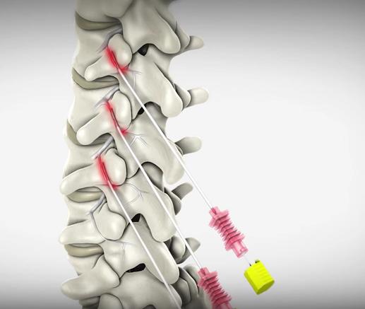
Cervical Facet radiofrequency neurotomy uses heat to create a lesion (damaged area) on the medial nerve. The lesion impairs the medial nerve’s ability to transmit signals about facet joint pain. Because the nerve is “turned off,” pain is not felt.
A cervical facet radiofrequency neurotomy is an outpatient procedure. You will wear a gown for the procedure and be positioned lying face down on a table. You will receive relaxation medicine before your procedure begins. The back of your neck will be sterilized and numbed with an anesthetic medication.
Your doctor will use a live X-ray image (fluoroscopy) to carefully insert and guide a needle-like tube (cannula) to the affected medial nerve. A small needle-like electrode (radiofrequency electrode) is inserted through the cannula. To ensure the cannula is in the correct position, a very mild electrical current is delivered through the electrode to the nerve. The nerve will briefly conduct pain signals and cause a muscle twitch, confirming that the correct nerve is targeted. Next, numbing medication is provided to the nerve in preparation for the treatment. Heat is delivered through the electrode to the nerve. The heat creates a lesion on the nerve. The heat disrupts the nerve’s ability to send signals about pain. At the end of the procedure, the cannula and electrode are removed. The process can be repeated for additional nerves that require treatment.


